How can we help you?
Clinical Excellence
Evidence-based care through innovation, advanced technology, and qualified professionals, ensuring exceptional patient outcomes
Excellent Patient Care
Compassionate, personalized, holistic approach leading to superior patient care
Transparent and Ethical
Upholding honesty and integrity in all interactions, ensuring trust through clear communication and ethical practices
Modern Infrastructure
Equipped with state-of-the-art facilities and cutting-edge technology to support superior patient care and innovative treatments
Why Choose Sterling
Why Choose
Sterling
Choose Sterling Hospitals, Gujarat's first NABH-accredited private hospital with over 20 years of excellence in patient care. Our 6 hospitals across the state offer comprehensive, tailored healthcare, touching lakhs of lives annually. Conveniently located and managed by compassionate professionals, Sterling Hospitals ensures accessible, top-tier care for every patient.
Our Specialities
Our
Specialities
From routine check-ups to specialized treatments, our dedicated team of healthcare professionals offers personalized care to meet all your needs on your journey to wellness.
Our Hospitals
Our
Hospitals
Checkout Sterling Hospitals near you to give a quick visit and get your checkup done by our best professionals


Transforming Lives with Expert Care
Watch inspiring recovery stories from our patients and explore our comprehensive healthcare services
Patient Success Stories
Real recovery journeys from our patients
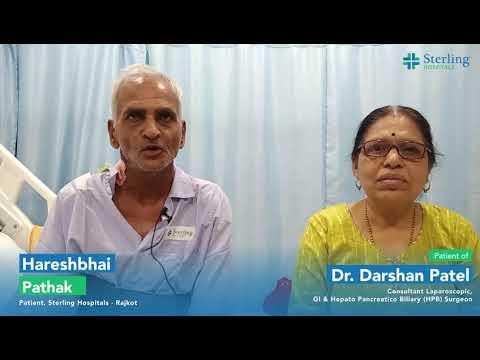
Expert Pancreatitis Care at Sterling Hospitals
Hareshbhai's health improved under the expert care of Dr. Darshan and the team at Sterling Hospitals.

Advanced Paediatric Oncology Care at Sterling Hospitals
Sterling Hospitals delivered advanced paediatric oncology care, preserving a young girl's liver with precision surgery

Treatment of Advanced Lung Cancer Complication at Sterling Hospitals, Rajkot
A life-saving Thoracoscopic Decortication successfully treated a lung cancer
Health Blogs
Health
Blogs
Explore Further: Checkout and Dive into more Blogs and keep yourself updated

Antacids are commonly used to relieve acid reflux, heartburn, and indigestion by neutralizing stomach acid. While these medications provide quick relief for many people, recent studies suggest a potential link between the frequent use of antacids and an increased risk of migraine attacks or severe headaches.
Though not all individuals who use antacids will experience headaches, there is growing evidence that certain ingredients in these medications, such as magnesium and calcium, can trigger or exacerbate headaches in susceptible individuals. Additionally, long-term use of antacids may disrupt the balance of gut bacteria, which has been implicated in gastrointestinal and neurological health.
Understanding this connection is paramount for those who suffer from migraines or chronic headaches, as making simple adjustments to diet or treatment options can potentially help reduce headache frequency and severity. This article explores how antacids might contribute to migraine attacks and provides alternative strategies for managing acid reflux.
How Are Acid Reflux and Migraine Episodes Connected?
The connection between acid reflux (GERD) and migraines is not immediately apparent. Still, research suggests that there may be a link between the two conditions, particularly when it comes to triggering or exacerbating migraine episodes. Several factors may contribute to this connection:
- Shared Triggers
- Both acid reflux and migraines can be triggered by similar factors, such as certain foods, stress, and lack of sleep. Foods like chocolate, citrus, spicy dishes, and caffeine can irritate the stomach, leading to acid reflux while also being common migraine triggers. This overlap means that individuals with both conditions may experience frequent flare-ups of both acid reflux and headaches.
- Gastrointestinal Disruption and Nervous System Interaction
- The gut & brain are closely linked through the gut-brain axis, a communication pathway between the gastrointestinal and central nervous systems. Disruptions in the digestive system, such as acid reflux, may send signals to the brain that can influence headache pathways. This connection could make individuals with acid reflux more prone to migraines or cluster headaches.
- Acid Reflux Medication and Migraine Risk
- Certain medications used to treat acid reflux, particularly antacids or proton pump inhibitors (PPIs), can also play a role in triggering or worsening migraines. Studies have shown that some individuals experience increased headache frequency when using antacids containing magnesium or calcium. These minerals, when taken in excess, can cause a fluctuation in neurotransmitters and blood flow to the brain, potentially triggering a migraine.
- Inflammation and Sensitivity
- Both GERD and migraines are associated with increased inflammation in the body. Chronic inflammation can heighten sensitivity in various areas, including the digestive system and the brain. The irritation caused by acid reflux can increase overall sensitivity, potentially making individuals more susceptible to migraines.
- Esophageal and Neurological Sensitivity
- The nerves involved in esophageal function, particularly the vagus nerve, can also impact migraine sensitivity. This nerve plays a key role in controlling stomach acid and digestive processes but can also influence pain pathways in the brain. In some individuals, overstimulation of this nerve from acid reflux may increase the likelihood of migraine episodes.
Why Proton Pump Inhibitors (PPIs) Are Associated with Migraine Episodes
Proton pump inhibitors (PPIs), commonly used to treat acid reflux and GERD by reducing stomach acid production, are associated with a boosted risk of migraine episodes in some individuals. This link is primarily due to PPIs' effects on the balance of certain minerals and neurotransmitters, which can influence headache pathways in the brain.
PPIs can alter the levels of magnesium and calcium in the body—two essential minerals that play a humongous role in muscle function, nerve signaling, & blood flow. Low magnesium levels are a common trigger for migraines, as they can affect blood vessel dilation and neuronal excitability.
Additionally, PPIs may disrupt gut bacteria and gut-brain communication, further exacerbating headache sensitivity. While not everyone using PPIs will experience migraines, individuals prone to headaches may need to monitor their symptoms and consider alternative treatments if they notice a correlation between PPIs and increased headache frequency.
When to See a Doctor: Recognizing the Need for Medical Guidance
Suppose you regularly experience acid reflux or migraines. In that case, monitoring how your symptoms interact and whether treatments like antacids or proton pump inhibitors (PPIs) may contribute to your headaches is essential.
If you notice a pattern where your migraines worsen after taking these medications or if your acid reflux symptoms are not adequately controlled, it's time to consult a healthcare professional. Your healthcare provider or a doctor can help identify potential triggers and recommend alternative treatments that better suit your needs.
Additionally, if you experience severe or chronic migraines, difficulty swallowing, unexplained weight loss, or other unusual symptoms, seeking medical advice is essential. Early help can help prevent complications and improve your quality of life by addressing your gastrointestinal and headache issues with a tailored, more effective treatment plan.
Sterling Hospital is Your Partner in Better Health Across Gujarat
At Sterling Hospital, we provide you with the utmost care and comfort throughout your journey to a healthier life. If you are looking for the best gastro surgeon in Ahmedabad, Vadodara, or Rajkot, we've got you. With considerable years of experience, we have built a team of the best gastro surgeons in these regions to provide you with premium treatment and the best results.
We have proven ourselves to be the best neurology hospital in Ahmedabad, Vadodara, Gandhidham, and Rajkot as well. We've got the best neurosurgeons in Rajkot, Vadodara, and Ahmedabad, who will be your partner throughout your journey. Contact Sterling Hospital to schedule a consultation and embark on a journey towards a healthier life.

GERD, or gastroesophageal reflux disease, is a persistent digestive issue where stomach acid or bile irritates the esophagus lining. This results in symptoms such as heartburn, regurgitation, & chest discomfort. This condition develops when the lower esophageal sphincter (LES), a muscle ring that typically blocks acid from returning to the esophagus, becomes weak or relaxed.
GERD is associated with a hiatal hernia very often. Here a portion of the stomach moves upward through the diaphragm into the chest area. This hernia can further exacerbate GERD symptoms by affecting the function of the LES. Both GERD and hiatal hernia are common, affecting millions of people worldwide.
While they can cause discomfort, with proper diagnosis and management—including lifestyle changes, medication, and, in some cases, surgery—the symptoms can be controlled, and further complications can be prevented.
The Link Between GERD and Hiatal Hernia
GERD and hiatal hernia often go hand in hand, with one condition frequently contributing to the other. While they are distinct issues, the presence of a hiatal hernia can significantly worsen GERD symptoms and vice versa. Here’s how they are connected:
- Hiatal Hernia Weakens the Lower Esophageal Sphincter (LES)
- The LES is a valve-like muscle that usually prevents stomach acid from flowing back in the esophagus. When a hiatal hernia occurs, part of the stomach moves into the chest cavity, causing the LES to malfunction. This allows stomach acid and food to flow back into the esophagus, leading to the signs and symptoms of GERD.
- Increased Pressure on the Stomach
- A hiatal hernia can also increase pressure within the stomach, which may push acid upward into the esophagus more easily. This added pressure can make GERD symptoms, such as heartburn and acid regurgitation, more frequent and severe.
- Worsened Symptoms
- People with both GERD and a hiatal hernia may experience intensified discomfort, including heartburn, chest pain, and difficulty swallowing. The hernia can make it harder for the LES to stay closed, leading to more frequent acid reflux episodes and prolonged irritation of the esophagus.
- Treatment Overlap
- Treating GERD and hiatal hernia often involves similar approaches, such as lifestyle modifications (diet changes, weight management, avoiding late-night eating), medications (antacids, proton pump inhibitors), and sometimes surgery. For patients with both conditions, treating the hiatal hernia may help reduce GERD symptoms, while controlling acid reflux can alleviate the impact of the hernia.
Diagnosing GERD and Hiatal Hernia
- Medical History and Symptom Review
- The first step in diagnosis involves discussing your symptoms with a doctor. They will ask about the frequency & intensity of symptoms like heartburn, chest pain, regurgitation, and difficulty swallowing. If you have a history of acid reflux or if lifestyle factors like diet, obesity, or smoking are contributing, this information can help guide further tests.
- Physical Exam
- While a physical exam alone cannot confirm GERD or a hiatal hernia, your doctor may check for signs of complications, such as difficulty swallowing or tenderness in the abdomen. This may help identify the severity of the condition.
- Upper Endoscopy (EGD)
- An upper endoscopy is often used to diagnose GERD and examine the esophagus, stomach, and duodenum. A flexible & thin tube with a camera in it (endoscope) is passed through the mouth to look for inflammation, ulcers, or other damage caused by acid reflux. In cases of GERD, it can also help identify a hiatal hernia.
- Barium Swallow X-ray
- In some cases, a barium swallow may be recommended. You’ll swallow a liquid that contains barium, which helps highlight the outline of your esophagus and stomach on an X-ray. This test can show if the stomach has pushed through the diaphragm, indicating a hiatal hernia, or if the esophagus is narrowing, which could suggest GERD.
- Esophageal Manometry
- This time, a thin tube is inserted through the nose into the esophagus, which records how well the LES works to prevent acid reflux. This test is instrumental when a doctor suspects abnormal esophageal function, which can be a factor in GERD and hiatal hernia.
- pH Monitoring
- To diagnose GERD precisely, pH monitoring involves inserting a tiny probe into the esophagus to measure the amount of acid refluxing into the esophagus over 24-48 hours. This test is instrumental in cases where symptoms don’t clearly match GERD but are still suggestive of acid reflux.
- Manometry and pH Monitoring Combined with Impedance Testing
- In complex cases where GERD symptoms don’t respond to treatment, impedance testing combined with manometry and pH monitoring may be used. This technique can evaluate both acid and non-acid reflux and provide a more comprehensive picture of esophageal function.
Treatment for GERD and Hiatal Hernia
- Lifestyle Modifications
- Dietary Changes: Avoid trigger foods like spicy foods, caffeine, chocolate, alcohol, and fatty meals, which can relax the lower esophageal sphincter (LES) & worsen reflux symptoms. Consuming smaller, more frequent meals & avoiding large meals before bedtime can also help.
- Weight Management: Being overweight puts additional pressure on the abdomen, which can worsen both GERD and a hiatal hernia. Losing & maintaining a healthy weight through diet and exercise can significantly reduce symptoms.
- Elevating the Head While Sleeping: Sleeping with your head raised on a pillow or adjustable bed can prevent acid from flowing back in the esophagus, particularly in individuals with GERD and hiatal hernia.
- Avoiding Tight Clothing: Tight-fitting clothes, especially around the waist, can boost pressure on the stomach and worsen symptoms. Loose-fitting attire is recommended.
- Surgical Interventions
- Fundoplication: A surgical procedure called fundoplication may be recommended for people with severe GERD that does not respond to medications or lifestyle changes. This surgery involves wrapping the stomach top around the LES, which strengthens it & prevents acid reflux.
- Laparoscopic Surgery: In some cases, surgery may be performed laparoscopically (minimally invasive) to repair a hiatal hernia. This procedure can help reposition the stomach and reduce symptoms by restoring the natural barrier between the stomach and esophagus.
- Endoscopic Procedures: Less invasive options, such as endoscopic treatments, may be considered for some individuals. These techniques use a scope to insert devices that can strengthen the LES or treat the hernia.
- Managing GERD and Hiatal Hernia Together
- Treatment often involves addressing both conditions simultaneously, as the two are interrelated. For example, managing the symptoms of GERD through diet, medication, and surgery can also help improve the symptoms of a hiatal hernia. Likewise, surgically correcting a hiatal hernia may alleviate GERD symptoms by restoring normal anatomy and function.
- Alternative and Complementary Therapies
- Herbal Remedies: Some individuals relieve GERD symptoms with herbal remedies like ginger, chamomile, or slippery elm. However, it's essential to consult a doctor before trying any sort of alternative treatments to ensure they are safe and effective.
- Acid-Alkaline Balance: While research on the alkaline diet is still limited, some people with GERD find that eating more alkaline foods (like leafy greens and root vegetables) and reducing acidic foods can help manage symptoms.
When to Seek Professional Help
While many people can manage GERD and hiatal hernia with lifestyle changes and medication, it's essential to consult your doctor if symptoms tend to be severe, persistent, or worsening. Chronic GERD can lead to severe complications such as esophageal ulcers, bleeding, or narrowing, while an untreated hiatal hernia may cause more severe pain or complications.
A tailored treatment plan that combines dietary adjustments, medications, and, when necessary, surgery can help alleviate discomfort & prevent long-term health issues associated with these conditions.
Sterling Hospital is Your Partner in Better Health Across Gujarat
At Sterling Hospital, we provide you with the utmost care and comfort throughout your journey to a healthier life. If you are looking for the best gastro surgeon in Ahmedabad, Vadodara, or Rajkot, we've got you. With considerable years of experience, we have built a team of the best gastro surgeons in these regions to provide you with premium treatment and the best results. Contact Sterling Hospital to schedule a consultation and embark on a journey towards a healthier future.

Hypertension, aka high blood pressure, occurs when the blood exerts an excessive amount of force against the walls of arteries over an extended period. This persistent elevation can harm blood vessels and organs, potentially resulting in various significant health complications.
Often referred to as a "silent killer" because it typically shows no immediate symptoms, hypertension can quietly damage the body for years before noticeable problems arise. If left untreated, it may lead to complications such as heart disease, kidney failure, Stroke, and vision loss.
Managing hypertension is crucial to prevent these severe outcomes, and while medication and lifestyle changes play a key role, early detection, and regular monitoring are essential in reducing the risks. Understanding the potential complications of hypertension is vital for anyone living with the condition, as it empowers patients to make informed decisions and protect their long-term health.
Hypertension and Its Link to Stroke: How High Blood Pressure Increases Your Risk
Hypertension is a grand contributor to the risk of Stroke, which ranks among the top causes of mortality and disability globally. Persistently high blood pressure increases the strain on arteries, reducing their flexibility and heightening the likelihood of damage.
This damage may cause blood clots to form or depleted blood vessels in the brain to rupture, either of which can trigger a stroke. There are two primary ways hypertension can increase stroke risk:
- Ischemic Stroke comes about when a blood clot blocks an artery leading to the brain. High blood pressure accelerates the process of atherosclerosis (hardening of the arteries), which causes plaque to build up and narrow the blood vessels. This makes it easier for a clot to form and block blood flow to the brain.
- Hemorrhagic Stroke: Chronic hypertension can also cause blood vessels to weaken and burst, leading to bleeding in the brain. This type of Stroke is more severe and can result in colossal damage to the brain or even death.
Hypertension and Its Connection to Metabolic Syndrome
Hypertension is often closely linked to metabolic syndrome, a cluster of risk factors that promote the likelihood of developing Stroke, heart disease, and type 2 diabetes. When high blood pressure is combined with other metabolic abnormalities, it significantly raises the risk of serious health complications. Here's how hypertension fits into metabolic syndrome:
- Elevated Blood Pressure
- High blood pressure is one of the key components of metabolic syndrome. If the blood pressure is consistently higher than 130/85 mmHg, it increases the risk of heart disease and Stroke. Increased blood pressure strains the heart and arteries, promoting the development of atherosclerosis (narrowing and hardening of the arteries).
- Insulin Resistance
- Insulin resistance, another hallmark of metabolic syndrome, happens when the cells of the body become less responsive to insulin. This condition leads to an increase in blood sugar levels, and in combination with hypertension, it can significantly boost the risk of developing type 2 diabetes and cardiovascular diseases.
- Abdominal Obesity
- It is the excess fat around the waist is another feature of metabolic syndrome that often coexists with hypertension. Fat around the abdomen increases the production of inflammatory substances, raising blood pressure and contributing to heart disease.
- Elevated Blood Sugar
- High blood sugar levels, often linked to insulin resistance, further contribute to hypertension. Elevated glucose levels can damage blood vessels, worsening the effects of high blood pressure and increasing the risk of kidney disease & heart failure.
- Abnormal Cholesterol Levels
- Metabolic syndrome often includes abnormal cholesterol levels, such as high triglycerides and low HDL (good) cholesterol. These changes, combined with high blood pressure, can increase the risk of cardiovascular diseases—heart attacks & strokes.
How Hypertension Can Damage Your Kidneys
- Increased Pressure on Blood Vessels
- The kidneys contain a vast network of blood vessels that filter waste & excess fluid from the blood. High blood pressure strains these vessels, leading to their damage. This can damage the kidneys' ability to filter waste efficiently.
- Narrowing and Hardening of Arteries (Atherosclerosis)
- Hypertension accelerates the process of atherosclerosis, where the arteries supplying the kidneys become narrower and less flexible due to plaque buildup. This reduces blood flow to the kidneys, depriving them of the oxygen and nutrients needed for proper function.
- Damage to Glomeruli
- The glomeruli are tiny filtration units within the kidneys. High blood pressure can damage these structures, leading to protein leakage into the urine (proteinuria), a sign of kidney dysfunction. Over time, this damage can worsen and lead to kidney failure.
- Decreased Kidney Function
- Damage to blood vessels in the kidneys degrades their ability to filter waste & regulate fluid balance effectively. This disruption can cause toxins and waste products to accumulate in the body, ultimately reducing kidney function.
- Kidney Enlargement
- Chronic high blood pressure can cause the kidneys to enlarge, which often leads to further damage and loss of kidney function. This condition can be detected in imaging tests and is a sign of ongoing kidney stress.
- Chronic Kidney Disease (CKD)
- Prolonged hypertension is a major contributor to the development of chronic kidney disease (CKD). Over time, as kidney function continues to decline, CKD can progress to kidney failure over time, requiring dialysis or even a kidney transplant.
Hypertension and Peripheral Artery Disease (PAD)
Peripheral artery disease (PAD) occurs when blood vessels outside the heart and brain, especially in the legs, become narrowed or obstructed. High blood pressure significantly contributes to PAD by promoting plaque buildup in the arteries, a condition known as atherosclerosis.
Chronic hypertension damages the arterial walls, triggering inflammation and the formation of fatty deposits over time. As the arteries become more constricted, blood flow to the legs is reduced, causing symptoms like leg pain, cramping, and weakness, especially when walking or exercising.
In severe cases, PAD can lead to ulcers, infections, or even tissue death, requiring more invasive treatments such as surgery or amputation. Managing hypertension is essential in reducing the risk of PAD and improving overall vascular health.
Hypertension and Vision Loss
High blood pressure can sometimes also lead to vision problems, including partial or complete vision loss, by impairing the delicate blood vessels in the eyes. Hypertension can lead to hypertensive retinopathy, which impacts the retina, the eye's structure responsible for dispatching visual signals to the brain.
High blood pressure can debilitate the blood vessels in the eye, causing them to leak & potentially resulting in blurred vision, swelling, or bleeding. Over time, untreated hypertension can cause more severe eye damage, leading to macular degeneration or even blindness. Regular eye exams are fundamental for detecting signs of hypertension-related vision loss early, allowing for intervention to preserve vision and prevent further damage.
Hypertension and Sexual Dysfunction
Hypertension is a common cause of sexual dysfunction in both men and women. In men, elevated blood pressure can impair blood vessels, limiting circulation to the penis and making it difficult to achieve or sometimes sustain an erection.
This condition is called erectile dysfunction (ED). In women, hypertension can impair blood flow to the genital area, leading to reduced sexual arousal and difficulty achieving orgasm. Managing hypertension through lifestyle changes, proper medication, and regular monitoring can help reduce the impact on sexual health, improving overall quality of life and preventing long-term issues.
Preventing the Complications of Hypertension: Key Steps for a Healthy Life
Here are some essential steps to avoid complications of hypertension:
- Monitor Your Blood Pressure Regularly
- Keep track of your blood pressure at home and during medical visits. Early detection of elevated blood pressure allows for prompt action to manage it before complications arise.
- Follow a Healthy Diet
- Adopt a heart-friendly diet that includes plenty of fruits, vegetables, whole grains, lean proteins, and essential minerals. Reduce consumption of sodium, processed foods, and unhealthy fats. The DASH diet, aka Dietary Approaches to Stop Hypertension, is particularly effective for controlling blood pressure.
- Maintain a Healthy Weight
- Losing even a bit of weight can remarkably lower blood pressure & lowers the risk of complications. Aim for a balanced diet combined with regular exercise.
- Exercise Regularly
- Physical activity helps lower blood pressure and strengthens the heart. Do at least 30 minutes of moderate-intensity exercise most days of the week, such as walking, cycling, or swimming.
- Limit Alcohol Intake
- Limit your alcohol infusion to one drink per day for women and two for men.
- Quit Smoking
- Quitting smoking improves cardiovascular health and lowers the risk of hypertension complications.
- Manage Stress Effectively
- Chronic stress is often seen to contribute to higher blood pressure. practice more stress-reducing activities such as yoga, meditation, deep breathing exercises, journaling or other hobbies that help you relax and unwind.
- Limit Caffeine Intake
- High amounts of caffeine can temporarily raise blood pressure in some people. Monitor your caffeine consumption and opt for decaffeinated beverages when possible.
- Take Medication as Prescribed
- If prescribed medication for hypertension, follow your doctor's instructions carefully. Consistent medication use and regular checkups can keep your blood pressure within a safe range and prevent long-term damage.
- Get Enough Sleep
- Poor sleeping habit is linked to higher blood pressure and other health problems. Get around 7-9 hours of quality sleep each night to help maintain a healthy blood pressure level.
- Stay Hydrated
- Drinking enough water helps maintain fluid balance in the body and supports overall health, including healthy blood pressure regulation.
- Regular Health Checkups
- Regular doctor visits and screenings for heart disease, kidney function, and other health issues are essential in detecting and addressing problems early, ensuring proper management of hypertension, & reducing the risk of complications.
Sterling Hospital is Your Partner in Heart Health Across Gujarat
At Sterling Hospital, we provide you with the utmost care and comfort throughout your journey to a healthier heart. If you are looking for the best cardiovascular surgeons in Ahmedabad, Vadodara, Rajkot, or Gandhidham, we've got you. With considerable years of experience, we have built a team of the best cardiologists in these regions to provide you with premium treatment and the best results. Contact Sterling Hospital to schedule a consultation and embark on a journey towards a healthier heart

A bone marrow transplant is a medical procedure where the diseased or damaged bone marrow is replaced with healthy blood-forming stem cells. Bone marrow is recognized as a spongy tissue inside bones that produces blood cells essential for carrying oxygen, fighting infections, and aiding in clotting.
When bone marrow fails to function correctly due to conditions like leukemia, lymphoma, or severe anemia, a transplant becomes a vital treatment option. The procedure involves infusing healthy stem cells into the patient's bloodstream, which then migrates to the bone marrow, & generating new, healthy blood cells.
This process can restore the body's ability to produce blood cells and bolster the immune system, offering hope for recovery & improved quality of life for individuals facing serious blood-related illnesses.
Types of Bone Marrow Transplants
Based on the source of stem cells, bone marrow transplants are categorized into three main types. Each type is suited to different medical conditions and individual patient needs.
- Autologous Transplant
- This type uses the patient's own stem cells. Before starting treatments like chemotherapy or radiation, stem cells are collected and stored.
- After treatment, the stored cells are reintroduced to the body to help recover the bone marrow's ability to produce healthy blood cells. Autologous transplants are often used for conditions like multiple myeloma or certain lymphomas.
- Allogeneic Transplant
- Stem cells are obtained from a person (donor) with a tissue type closely matching the patient's. A sibling or unrelated donor may be a match.
- Allogeneic transplants are common for conditions such as leukemia, severe immune system disorders, and some genetic diseases.
- Haploidentical Transplant
- This is a form of allogeneic transplant where the donor is a half-match, often a parent or sibling.
- It expands the pool of potential donors and is increasingly used in cases where a fully matched donor is unavailable. It is suitable for various blood cancers and immune deficiencies.
Why Bone Marrow Transplants Are Done
A bone marrow transplant is performed when the bone marrow itself cannot produce enough healthy blood cells, a condition that can arise due to diseases, treatments, or inherited conditions. It is a critical option for patients with life-threatening illnesses that affect the blood and immune system.
For individuals with blood cancers such as leukemia and lymphoma, a transplant can replace damaged or cancerous marrow with healthy stem cells. This helps restore the body's ability to produce normal blood cells and reduces the likelihood of disease relapse.
Patients undergoing high-dose of chemotherapy or radiation therapy for cancer often require a transplant. These treatments can destroy not only cancer cells but also healthy bone marrow, making a transplant essential for recovery.
In cases of non-cancerous conditions like aplastic anemia or severe immune system disorders, a transplant helps replenish defective or non-functioning bone marrow. Similarly, transplants can provide a curative option for genetic conditions, namely sickle cell anemia or thalassemia, by replacing faulty stem cells.
Ultimately, the procedure is a powerful intervention that aims to restore health, extend life, and provide a chance for recovery in the face of debilitating conditions.
Risks of Bone Marrow Transplant
While a bone marrow transplant can be life-saving, it carries significant risks. The risks depend on factors such as the patient’s overall health, age, the type of transplant, and the underlying medical condition. Here are the key risks associated with the procedure:
- Infections
- The immune system is weakened during and after the transplant, making patients highly susceptible to infections.
- Graft-versus-host disease (GVHD)
- In allogeneic transplants, the donor’s immune cells may attack the recipient’s tissues, leading to skin, liver, and digestive tract complications.
- Organ damage
- High-dose chemotherapy or radiation before the transplant can damage organs like the liver, heart, or lungs.
- Bleeding and anemia
- Low platelet counts during recovery can increase the risk of bleeding, while low red blood cell levels may cause anemia.
- Relapse of disease
- There is a possibility that the underlying condition, such as cancer, may return even after a transplant.
- Fertility issues
- The treatments involved can affect fertility, potentially leading to temporary or permanent infertility.
- Long-term side effects
- Some patients may experience chronic conditions such as cataracts, hormone imbalances, or secondary cancers later in life.
- Rejection of the transplant
- In rare cases, the body may sometimes reject the donor stem cells, leading to transplant failure.
Preparation for Bone Marrow Transplant
Preparation for a bone marrow transplant involves several steps to ensure the procedure’s success and minimize risks. It requires coordination between the patient, medical team, and caregivers. Here’s a detailed guide:
- Comprehensive Medical Evaluation
- Undergo detailed blood tests, imaging scans, and heart and lung function tests to confirm fitness for the transplant.
- Discuss the benefits & risks of the transplant with your doctor to set clear expectations.
- Finding a Suitable Donor
- If undergoing an allogeneic transplant, identify a donor through tissue typing. This could be a family member or an unrelated match from a donor registry.
- For autologous transplants, schedule stem cell collection and storage well in advance.
- Pre-transplant Conditioning
- Complete chemotherapy or radiation therapy as prescribed to destroy diseased bone marrow or cancer cells and suppress the immune system.
- Be prepared for potential side effects such as fatigue, nausea, or hair loss.
- Nutritional and Physical Health Optimization
- Maintain a healthy diet to build strength and resilience. A dietitian may recommend a plan rich in nutrients to support recovery.
- If possible, engage in light physical activity to keep the body in optimal condition.
- Psychological and Emotional Preparation
- Seek counseling or join a support group to address concerns and reduce anxiety about the procedure.
- Involve family and friends for emotional support and caregiving during the recovery period.
- Arrange for Post-Transplant Care
- Plan to stay near the transplant center, especially if follow-up visits and monitoring are required frequently.
- Arrange for caregivers who can assist with daily activities during the recovery phase.
- Preventive Measures Against Infections
- Complete necessary vaccinations before the procedure as your doctor has advised.
- Maintain strict hygiene and avoid crowded places to reduce exposure to infections.
- Understand Financial and Logistical Aspects
- Check your insurance coverage for transplant costs, including pre-and post-procedure expenses.
- Plan for time off work and discuss arrangements for managing household responsibilities.
- Packing Essentials for Hospital Stay
- Pack comfortable clothing, personal hygiene items, and entertainment to make the hospital stay more manageable.
- Include any prescribed medications and a list of current health conditions.
What to Expect During the Process of Bone Marrow Transplant
During the transplant, patients receive the stem cells through a procedure similar to a blood transfusion. It is typically painless and lasts a few hours. Before the transplant, a conditioning phase involving high-dose chemotherapy or radiation may be administered to prepare the body by eradicating diseased cells and suppressing the immune system.
Side effects like nausea, fatigue, or mouth sores are common during this phase. On the transplant day, the stem cells are infused through an IV line, and patients are closely monitored for any immediate reactions, such as fever or chills. The process is not invasive, but the body’s response can vary widely.
What to Expect After the Bone Marrow Transplant
Post-transplant recovery is a gradual process and requires close monitoring in a hospital or outpatient setting. The first few weeks are critical as the transplanted stem cells settle in the bone marrow of the new body and produce new blood cells, a process called engraftment.
During this time, patients may experience fatigue, an increased risk of infections & low blood cell counts. Regular blood tests, medications to prevent infections, and transfusions may be necessary. Long-term, patients may face side effects such as graft-versus-host disease (in allogeneic transplants) or organ complications.
Recovery timelines vary, with most patients regaining significant health within a few months to a year.
Diet and Lifestyle Factors After a Bone Marrow Transplant
A healthy and cautious lifestyle plays a vital role in recovery after a bone marrow transplant. Nutrition is one of the most critical aspects of helping the body rebuild strength & immunity.
A transplant often weakens the immune system, making dietary choices particularly important.
Dietary Considerations
- Go for a well-balanced diet rich in protein, vitamins, & minerals to promote healing and cell regeneration. Foods like lean meats, eggs, dairy products, and legumes are excellent protein sources.
- Incorporate plenty of fresh fruits and vegetables, but ensure they are thoroughly washed or cooked to reduce the risk of bacteria infections or other pathogens.
- Avoid raw or undercooked foods, including sushi, rare meats, or unpasteurized dairy, as these may harbor harmful microorganisms.
- Stay hydrated by drinking plenty of water. Try to avoid sugary or caffeinated beverages.
- Work with a dietitian to address specific dietary needs, especially if experiencing side effects like nausea or difficulty swallowing.
Lifestyle Factors
- Maintain strict hygiene by washing hands frequently, using sanitizers, and keeping living spaces clean.
- Limit exposure to crowds and sick individuals to minimize the risk of infections and diseases, especially during the first few months post-transplant.
- Follow a tailored exercise routine approved by your doctor to rebuild physical strength and improve overall energy levels.
- Ensure adequate rest to allow the body time to recover, balancing activity with periods of relaxation.
- Avoid smoking, alcohol, & other substances that can stress the immune system or impair organ function.
Results and Conclusion
A bone marrow transplant can be a transformative procedure, offering a chance for recovery and improved quality of life for individuals facing severe blood-related conditions. Success varies based on factors like the underlying disease, the patient’s overall health, & the type of transplant performed.
Many patients experience remission or significant improvement in their condition, regaining the ability to produce healthy blood cells and strengthening their immune system. However, the journey requires patience, as recovery may take months to a year, and long-term follow-ups are necessary to monitor progress and manage any complications.
Understanding the process, risks, and lifestyle adjustments is essential for patients and families to make informed decisions and ensure a smoother experience. With advancements in medical technology & personalized care, bone marrow transplants continue to provide hope and life-saving options for those in need. Patients can confidently navigate this challenging but potentially life-changing procedure by staying informed and prepared.
Sterling Hospital is Your Partner in Better Health Across Gujarat
Sterling Hospital has proven itself to be the best hospital of Haematology, Haemato-Oncology, and BMT in Ahmedabad - Gurukul, Ahmedabad - Sindhu Bhavan, and Rajkot. We provide our patients with the utmost care and comfort throughout their battle against their problems while striving to provide them with premium treatments and the best results. We've got the best doctors in Haemato-oncology, who will be your partner throughout your journey.
Contact Sterling Hospital to schedule a consultation and embark on a journey towards a healthier life.

A stroke comes about when blood flow to a part of the brain is either reduced or interrupted, preventing brain tissue from receiving essential oxygen and nutrients. In a few minutes, brain cells begin to die, leading to potential brain damage, disability, or death.
There are two preliminary types of strokes: ischemic & hemorrhagic. Ischemic strokes, the most common type, result from narrowing or blockages of the arteries supplying blood to the brain, often due to blood clots or fatty deposits.
Hemorrhagic strokes appear when a blood vessel in the brain bursts, causing bleeding around or into the brain tissue. Recognizing stroke symptoms—such as sudden numbness or weakness in the face, leg or arm, especially on one side of the body; vision problems; dizziness; confusion; trouble speaking or understanding speech; or severe headache—is crucial, as prompt medical treatment can significantly improve outcomes.
The Link Between Stress and Stroke
Stress is considered to be a natural reaction to challenges, but chronic stress can profoundly impact the body, including the cardiovascular system. Over time, persistent stress may increase the risk of stroke.
Stress triggers the release of some hormones such as cortisol & adrenaline, which is responsible for preparing the body for a “fight or flight” response. While this reaction is helpful in short bursts, prolonged exposure can strain the heart and blood vessels.
Chronic stress often leads to unhealthy habits such as overeating, smoking, or inactivity, which can raise blood pressure and cholesterol—key risk factors for stroke. Additionally, stress can directly cause physical changes in blood vessels, potentially contributing to blockages or ruptures.
For those already at risk due to conditions like high blood pressure or diabetes, unmanaged stress further amplifies the danger. Understanding & managing stress is not just about mental well-being but also about safeguarding long-term health.
Types of Stress That Can Influence Stroke Risk
Not all stress is the same, and different types can affect stroke risk in various ways. Here are the key categories:
- Acute Stress: Acute stress occurs during short-term events, like an argument, a deadline, or a sudden shock. Severe episodes of acute stress can also cause a brief spike in your blood pressure. It may also lead to arrhythmias, which may increase the likelihood of a stroke in high-risk individuals.
- Chronic Stress: Chronic stress stems from ongoing pressures, such as financial difficulties, work-related demands, or caregiving responsibilities. This persistent strain on the body can result in long-term inflammation, high blood pressure, and vascular damage, significantly elevating stroke risk.
- Emotional Stress: Feelings of grief, anxiety, or anger can place added strain on the cardiovascular system. Studies have shown that intense emotions can trigger transient ischemic attacks (TIAs) or even full strokes in vulnerable individuals.
- Physical Stress: Overexertion, illness, or injury can put the body under physical stress. For individuals with underlying conditions like atherosclerosis, these stressors can exacerbate the risk of clot formation or vessel rupture.
- Work-Related Stress: Job strain, characterized by high demands and low control over decisions, is a well-recognized contributor to hypertension and stroke. Prolonged exposure to such environments can create a cycle of unhealthy coping mechanisms and physiological stress.
Recognizing Symptoms of Stress-Induced Stroke
- Sudden Weakness or Numbness
- Weakness, numbness, or paralysis around the face, arm, or leg—especially on one side of the body—is a hallmark sign. A drooping face is often a clear indicator.
- Confusion or Trouble Speaking
- Difficulty speaking, slurred speech, or an inability to understand language can signal that the brain's communication centers are affected
- Vision Problems
- Double vision or sudden blurred in one or both eyes or complete loss of vision in one eye could indicate a stroke
- Severe Headache
- An intense headache, often described as the "worst headache ever," can be a warning sign, especially if it's accompanied by vomiting, dizziness, or altered consciousness
- Loss of Balance or Coordination
- Dizziness, loss of balance, or trouble walking might signal a stroke
- Fatigue or Mental Fog
- Prolonged stress might blur the line between mental exhaustion and a stroke symptom, but sudden, severe fatigue or mental fog should not be ignored
The acronym FAST (Face drooping, Arm weakness, Speech difficulties, Time to call for help) is a simple way to remember the key warning signs. Recognizing these symptoms early can make a life-saving difference.
Prevention: Reducing Stress and Stroke Risk
While stress cannot always be avoided, managing it effectively can significantly lower the risk of stroke. Here are practical steps to protect your health:
- Practice Stress Management
- Engage in relaxation techniques such as journaling, meditation, or yoga to calm the mind and body. Regular mindfulness practices can reduce cortisol levels and improve heart health.
- Adopt a Healthy Lifestyle
- Exercise Regularly: Physical activity reduces stress & improves cardiovascular health by lowering blood pressure & cholesterol levels. Do around 30 minutes of moderate exercise, if not daily, then most days.
- Eat a Balanced Diet: Include heart-healthy foods like fruits, vegetables, whole grains, lean protein, and healthy fats. Limit salt, sugar, and processed foods to control blood pressure and weight.
- Seek Social Support
- Build a strong support system of friends and family. Sharing concerns and seeking help during challenging times can alleviate emotional burdens.
- Avoid Unhealthy Coping Mechanisms
- Limit alcohol intake, quit smoking, and avoid overeating. These habits may temporarily reduce stress but can exacerbate long-term health risks.
- Monitor Health Regularly
- Keep track of your cholesterol, blood sugar levels, & blood pressure. Regular check-ups with your doctor can catch issues early and reduce stroke risk.
- Prioritize Sleep
- Quality sleep helps the body recover from stress and supports overall health. Strive to get at least 7–9 hours of uninterrupted sleep per night
- Manage Work-Related Stress
- Set boundaries, delegate tasks, and take regular breaks to avoid burnout. Finding a balance between work and personal life is essential for long-term health.
When to See a Doctor
Recognizing when to seek medical help is critical in managing stress & reducing the risk of stroke. Here's when you should consult a doctor:
- Chronic or Unmanageable Stress
- If stress persists despite efforts to manage it or begins to interfere with your daily life, it's Time to consult a healthcare provider. Symptoms such as persistent anxiety, irritability, or trouble sleeping may indicate the need for professional support.
- Signs of High Blood Pressure
- Stress often contributes to high blood pressure, a significant risk factor for stroke. Regular monitoring is essential, and a doctor can provide medication or lifestyle recommendations if your readings consistently exceed expected levels.
- Symptoms of a Stroke
- Numbness or weakness in the areas of face, arm, or leg (especially on one side).
- Difficulty speaking or understanding speech.
- Vision problems, severe headaches, or dizziness.
- Time is crucial during a stroke, and prompt treatment can prevent severe complications.
- Unusual Physical Symptoms Related to Stress
- If stress leads to persistent headaches, chest pain, irregular heartbeats, or other unusual symptoms, a doctor can rule out underlying conditions and provide treatment.
- Difficulty Coping with Emotional Stress
- If you feel overwhelmed by grief, depression, or anxiety, mental health professionals can offer counseling or recommend other therapies to manage emotional stress effectively.
Sterling Hospital is Your Partner in Better Health Across Gujarat
Advances in mental healthcare have empowered us with effective treatments, significantly improving the lives of those facing mental health disorders. We provide access to these latest treatments through our team of top psychiatrists and psychologists in Vadodara and Rajkot.
Remember, taking that first step towards your mental well-being begins with you. Acknowledging the need for support and seeking professional guidance is key. Don't hesitate to reach out. Contact Sterling Hospital today to schedule a consultation and embark on a journey towards improved mental health.

Alzheimer's disease is a progressive neurological disorder that gradually impairs memory, thinking, and behavior. It affects millions of people worldwide, making it one of the most common forms of dementia.
While the disease typically impacts older adults, it’s important to recognize its symptoms early, understand the condition, and know what to expect. This article provides an overview of Alzheimer’s disease, aimed at helping patients and their families navigate the challenges that come with it.
Symptoms of Alzheimer's Disease
Alzheimer’s disease manifests in various ways as it progresses. These symptoms often worsen over time and impact different aspects of daily life.
- Memory Loss: One of the earliest and most recognized signs of Alzheimer's is forgetfulness. Initially, patients may struggle to remember recent events, names, or conversations. As the disease progresses, they may forget important personal details, such as their address or the names of loved ones.
- Thinking and Reasoning Difficulties: Alzheimer's patients often experience difficulty concentrating and performing tasks that require logic and problem-solving. Simple calculations, making sense of time, and even basic decision-making can become challenging.
- Difficulty Making Decisions and Planning: Everyday activities such as managing finances, keeping track of appointments, or preparing a meal become overwhelming as Alzheimer's progresses. This can lead to frustration for both the individual and their caregivers.
- Challenges in Performing Familiar Tasks: People with Alzheimer’s may find it hard to complete tasks they’ve done many times before, such as driving to a familiar location or following a recipe. The disease interrupts the brain’s ability to sequence actions, making even routine activities difficult.
- Behavioral and Personality Changes: Alzheimer’s disease can cause significant changes in mood and personality. Individuals may become anxious, suspicious, or withdrawn. Irritability, depression, and mood swings are also common as the disease advances. In some cases, patients may experience hallucinations or delusions.
Diagnosis of Alzheimer's Disease
Diagnosing Alzheimer’s involves a comprehensive evaluation that looks at several aspects of a patient's health and behavior. The diagnostic process generally includes:
- Medical History and Symptom Review: The doctor will ask about any memory issues, changes in behavior, or difficulties with daily activities. They’ll also look into the patient’s medical history, family history of dementia, and any existing health conditions.
- Cognitive and Neuropsychological Tests: To assess memory, thinking, and problem-solving abilities, patients undergo cognitive tests that measure functions like memory recall, language skills, and the ability to follow instructions. These tests help doctors pinpoint whether cognitive decline is related to Alzheimer's or another condition.
- Physical and Neurological Exams: A physical exam helps rule out other potential causes of dementia, such as thyroid disorders or vitamin deficiencies. Neurological exams, including checking reflexes, muscle tone, and coordination, assess overall brain function.
- Mental Health Assessments: Depression and anxiety are common in Alzheimer's patients and may sometimes mask cognitive decline. Mental health evaluations help doctors differentiate between Alzheimer’s and other psychological conditions.
Diagnostic Tests for Alzheimer's Disease
To further refine the diagnosis and rule out other conditions, a series of tests are often conducted, which may include:
- Brain Imaging: Advanced imaging techniques, such as MRI (Magnetic Resonance Imaging) and CT (Computed Tomography) scans, are used to rule out brain tumors, strokes, or other structural abnormalities. These scans can also reveal brain atrophy, a common feature in Alzheimer’s patients.
- PET Scan (Positron Emission Tomography): PET scans help doctors visualize amyloid plaques, which are abnormal clumps of protein often associated with Alzheimer's disease. This imaging test is especially useful for diagnosing Alzheimer's in its early stages.
- Blood and Spinal Fluid Tests: Researchers are exploring the use of blood tests to detect biomarkers associated with Alzheimer’s, such as beta-amyloid and tau proteins. In some cases, a lumbar puncture (spinal tap) is used to examine cerebrospinal fluid for signs of these proteins, which are indicative of Alzheimer's.
Treatment Options for Alzheimer's Disease
Currently, there is no cure for Alzheimer’s disease, but treatments can help manage symptoms and slow progression. Treatment plans are typically tailored to each individual and may include:
- Medications to Manage Symptoms: Several drugs are approved to treat the cognitive symptoms of Alzheimer's, including Cholinesterase Inhibitors & Memantine These medications do not stop the disease but may help manage symptoms for a time.
- Behavioral and Psychological Symptom Management: As Alzheimer's progresses, patients may experience mood swings, anxiety, or aggression. Medications such as antidepressants or antipsychotics may be prescribed to address these issues, but their use is carefully monitored due to potential side effects.
- Lifestyle Modifications and Cognitive Rehabilitation: Non-drug therapies can play a crucial role in managing Alzheimer's. Cognitive rehabilitation programs help patients maintain mental function, and lifestyle changes, such as regular exercise, a healthy diet, and engaging in mental and social activities, may help slow the disease's progression.
- Supportive Therapies: Various therapies are available to improve the quality of life for Alzheimer's patients, including:
- Occupational Therapy to help patients maintain independence in daily activities.
- Speech and Language Therapy for those experiencing difficulty with communication.
- Support for Caregivers: Alzheimer’s disease doesn’t only affect the patient; it profoundly impacts family members and caregivers. Support groups, respite care services, and counseling can provide essential assistance to those caring for individuals with Alzheimer’s.
Safety Considerations for Alzheimer’s Patients
Ensuring safety is one of the most important aspects of caring for someone with Alzheimer's. Because the disease affects memory, judgment, and motor skills, individuals may face risks like falls, wandering, and accidental injuries. Here’s how to address these risks:
- Preventing Falls: Alzheimer's patients are at an increased risk of falling due to poor balance, confusion, or impaired vision. To prevent falls:
- Remove tripping hazards like loose rugs, electrical cords, and clutter.
- Install handrails in hallways, bathrooms, and staircases.
- Ensure rooms are well-lit, particularly at night, by installing motion-activated lights.
- Consider using non-slip mats in bathrooms and other areas prone to moisture.
- Wandering Prevention: Many people with Alzheimer's experience disorientation and may wander away from home, putting them in dangerous situations. To reduce wandering:
- Install locks on doors and windows that are difficult for patients to operate.
- Use motion sensors or door alarms to alert caregivers if the patient attempts to leave the house.
- Create a secure outdoor space where they can walk safely without leaving the premises.
- Consider a GPS tracking device or wearable ID bracelet so that if a person does wander, they can be quickly located.
- Safe Navigation: Alzheimer's patients can become easily confused or disoriented, making it hard to navigate even familiar environments. Helpful measures include:
- Labeling doors and cupboards with pictures or simple words to help patients find items more easily.
- Using color contrasts in floor and wall surfaces to help patients distinguish between rooms.
- Keeping frequently used items like keys, glasses, or a favorite chair in the same place every day to create familiarity and reduce confusion.
Promoting Independence and Reducing Anxiety
Supporting the independence of Alzheimer’s patients in a safe way is essential for their dignity and self-esteem. There are several ways to adjust the home environment to allow patients to maintain as much independence as possible:
- Simplify Daily Tasks: Break down complex activities, such as getting dressed or making a cup of tea, into simpler steps. Place easy-to-use items where they are most needed, such as setting out clothing in the order it should be put on.
- Create Routines: Consistency is key for reducing anxiety and confusion. Establish regular daily routines, including meal times, bathing, and sleep schedules, to create a sense of structure.
- Use Familiar Objects: Surrounding Alzheimer’s patients with familiar objects—like family photos, personal mementos, or cherished furniture—can help them feel more grounded and reduce feelings of disorientation.
- Limit Overstimulation: Alzheimer's patients can become easily overwhelmed by noise, large crowds, or even clutter. Maintain a calm and quiet home environment by reducing background noise (like TVs or radios) and limiting large gatherings in the home. Providing a quiet, comfortable space for rest or relaxation can help reduce anxiety.
Emotional and Behavioral Support
Caring for someone with Alzheimer's also involves providing emotional and behavioral support, as patients may experience mood swings, frustration, or aggression as part of their illness. Here’s how to create an emotionally supportive environment:
- Provide Reassurance and Comfort: Alzheimer’s patients often feel confused, anxious, or frightened, especially when they struggle to remember or complete tasks. Offering gentle reassurance, physical touch like a hand on their shoulder, and a calm tone of voice can help soothe their anxieties.
- Be Patient and Flexible: While it’s essential to establish a routine, be flexible when needed. Alzheimer's patients may resist certain activities or forget steps in their daily routine, so being patient and adapting to their changing needs is important.
- Engage in Meaningful Activities: Engaging Alzheimer’s patients in simple, meaningful activities can help reduce agitation and give them a sense of purpose. Activities such as listening to music, folding towels, watering plants, or working on a jigsaw puzzle can provide mental stimulation and emotional satisfaction.
Adapting the Environment Over Time
Alzheimer’s is a progressive disease, and the needs of the individual will change as the disease advances. Regularly reassessing the home environment and making adjustments can ensure it remains a safe and supportive space.
- Adjustments to the Bedroom: As Alzheimer's progresses, patients may have difficulty sleeping, which can lead to restlessness at night. To improve sleep quality:
- Ensure the bedroom is quiet, comfortable, and free of bright lights.
- Remove mirrors, as some patients become confused by their reflection.
- Use bed rails or place a mattress on the floor if falls from bed are a concern.
- Supervision and Professional Support: In the later stages of the disease, closer supervision may be necessary to ensure the patient's safety. Family members may want to consider hiring professional caregivers or exploring respite care services to provide additional support.
Sterling Hospital is Your Partner in Better Health Across Gujarat
Sterling Hospital has proven itself to be the best neurology hospital in Ahmedabad, Vadodara, Gandhidham, and Rajkot. We provide our patients with the utmost care and comfort throughout their battle against their neurological problems while striving to provide them with premium treatments and the best results. We've got the best neurosurgeons in Rajkot, Vadodara, and Ahmedabad, who will be your partner throughout your journey. Contact Sterling Hospital to schedule a consultation and embark on a journey towards a healthier life.

Heart disease describes various conditions affecting the heart's structure and function. It is a leading cause of death worldwide, but it can often be prevented or at least managed through lifestyle changes and medical treatment.
Understanding heart disease is crucial for patients and the general public to make informed decisions about their health. Here's a detailed overview to help you navigate this complex topic.
What is Heart Disease?
Heart disease encompasses several conditions that affect the heart, including:
- Coronary artery disease (CAD): A common type of plaque accumulates in the arteries that deliver blood to the heart. This buildup can restrict blood flow, potentially causing angina, heart attacks, or even heart failure.
- Arrhythmias: Irregular heartbeats that range from harmless to life-threatening. Common types include atrial fibrillation & ventricular tachycardia.
- Heart valve disease: Heart valve dysfunction, where one or more valves fail to function correctly, can disrupt the normal flow of blood through the heart.
- Heart failure: Despite the name, this doesn't mean the heart stops. Instead, it indicates the heart isn't pumping blood as effectively as it should.
- Congenital heart defects: These are heart problems present at birth and can affect the heart's structure, leading to issues in how blood flows through it.
- Cardiomyopathy: A disease caused by the heart muscle that can lead to heart failure.
Symptoms of Heart Disease
Symptoms may vary depending on the type of heart disease you have. Common signs to watch out for include:
- Chest pain or discomfort (angina), especially during physical activity.
- Shortness of breath.
- Fatigue or weakness.
- Palpitations or irregular heartbeat.
- Swelling in the legs, ankles, or feet (edema).
- Fainting or dizziness.
Causes and Risk Factors
The causes of heart disease are multifactorial, involving a blend of genetic, lifestyle, and environmental factors.
- Age: Risk increases with age.
- High blood pressure: Extra strain on the heart and arteries.
- High cholesterol: Plaque builds up in the arteries, increasing the risk of CAD and heart attacks.
- Smoking: Damages the blood vessels.
- Diabetes: High blood sugar can damage the heart and blood vessels.
- Obesity: Extra weight forces the heart to work harder, raising the risk of heart failure, CAD, and high blood pressure.
- Inadequate physical activity: Contributes to an elevated likelihood of developing conditions such as hypertension (high blood pressure), high cholesterol, and diabetes.
- Suboptimal nutrition: Diets rich in saturated and trans fats, sodium (salt), and refined sugars are associated with an increased susceptibility to cardiovascular disease.
- Genetic predisposition/Family background: Having a family history of heart disease indicates a potentially higher individual risk.
- Biological sex: Men generally face a greater risk earlier in life, whereas the risk for women tends to increase following the onset of menopause.
Diagnosing Heart Disease
- Electrocardiogram (ECG or EKG): This test records your heart's electrical activity and can detect arrhythmias and other abnormalities.
- Echocardiogram: It is an ultrasound of the heart that shows its structure and function, helping diagnose valve disease and heart failure.
- Stress test: Measures how your heart responds to exertion, often used to detect CAD.
- Cardiac catheterization: Involves threading a thin tube through the blood vessels to the heart to check for blockages in the coronary arteries.
- Blood tests: These can measure cholesterol levels, markers of inflammation, and enzymes that indicate a heart attack.
Treatment Options
Treatment depends on the type and severity of heart disease but often involves a combination of lifestyle changes, medications, and sometimes surgical interventions:
- Lifestyle changes: A heart-healthy diet, exercise, and quitting smoking are crucial to managing many types of heart disease.
- Medications may include drugs to lower cholesterol, control blood pressure, or prevent blood clots. For arrhythmias, antiarrhythmic medications can be prescribed.
- Surgical procedures: For severe cases, surgeries like angioplasty, stent placement, coronary artery bypass grafting (CABG), valve repair or replacement, or heart transplantation may be necessary.
Living with Heart Disease
For many, living with heart disease involves ongoing management. Regular check-ups with a cardiologist, adhering to prescribed medications, and following lifestyle recommendations are critical to preventing further complications. Familiarizing yourself with your condition and learning about new treatments and technologies that may benefit you is also essential.
Preventing Heart Disease
Some factors, like age and genetics, cannot be controlled, but many can be mitigated through lifestyle changes. Here are some strategies to help prevent heart disease:
- Exercise regularly
- Eat a heart-healthy diet
- Maintain a healthy weight
- Quit smoking
- Manage stress
- Control blood pressure and cholesterol levels
- Limit alcohol consumption
Sterling Hospital is Your Partner in Heart Health Across Gujarat
At Sterling Hospital, we provide you with the utmost care and comfort throughout your journey to a healthier heart. If you are looking for the best cardiovascular surgeons in Ahmedabad, Vadodara, Rajkot, or Gandhidham, we've got you. With considerable years of experience, we have built a team of the best cardiologists in these regions to provide you with premium treatment and the best results. Contact Sterling Hospital to schedule a consultation and embark on a journey towards a healthier heart.

It is a common and dangerous misconception that only heavy drinkers develop liver cirrhosis. In reality, the causes of this severe condition are far more diverse, affecting millions worldwide regardless of their history with alcohol.
Liver cirrhosis is a progressive and irreversible disease where healthy, functional liver tissue is gradually replaced by non-functional scar tissue (fibrosis). This scarring disrupts the liver's architecture, impeding the normal flow of blood and severely impairing its ability to perform essential, life-sustaining functions. While existing scar tissue cannot be reversed, timely diagnosis and intervention are critical to halting or slowing the progression of further damage and preventing life-threatening complications.
The Liver’s Essential Role
The liver is one of the body’s largest and most vital organs, acting as a powerhouse for hundreds of functions, including:
- Detoxification: Filtering toxins, drugs, and waste products from the blood.
- Metabolism: Processing fats, carbohydrates, and proteins from digested food.
- Production: Manufacturing crucial proteins, clotting factors, and bile for digestion.
- Storage: Storing vitamins, minerals, and glucose (as glycogen) for energy.
When cirrhosis develops, these functions are compromised, leading to systemic health problems.
Causes of Cirrhosis: A Diverse Spectrum
While alcohol abuse is a well-known risk factor, the majority of current cirrhosis cases stem from other chronic conditions.
- Chronic Alcohol Abuse: Long-term, excessive consumption of alcohol leads to alcoholic hepatitis, which progresses to cirrhosis.
- Viral Hepatitis: Chronic infections with Hepatitis B or Hepatitis C viruses are major global causes. These viruses are spread through exposure to infected blood, sexual contact, or from mother to child. Treatment with antiviral medications can often suppress Hepatitis B and cure Hepatitis C, preventing progression to cirrhosis.
- Metabolic Dysfunction-associated Steatotic Liver Disease (MASLD) and MASH: This category represents the fastest-growing cause of liver disease globally. MASLD (formerly NAFLD) involves excess fat buildup in the liver. When this fat causes inflammation and cell damage, it is called Metabolic Dysfunction-associated Steatohepatitis (MASH) (formerly NASH). MASH is strongly linked to Metabolic Syndrome—a cluster of conditions including obesity, Type 2 diabetes, high cholesterol, and high blood pressure—and is a common pathway to cirrhosis.
- Chronic Bile Duct Disorders: Conditions that damage or block the small bile ducts, causing bile to back up and injure liver cells. Examples include:
- Primary Biliary Cholangitis (PBC): An autoimmune disorder that slowly destroys the small bile ducts.
- Primary Sclerosing Cholangitis (PSC): Inflammation and scarring of the large bile ducts.
- Genetic and Metabolic Conditions:
- Hemochromatosis: An inherited disorder leading to excessive iron buildup in the liver and other organs.
- Wilson’s Disease: A rare, inherited disorder causing copper accumulation in the body.
- Autoimmune Hepatitis: A condition where the body’s own immune system mistakenly attacks and inflames the liver.
- Toxic Hepatitis: Long-term exposure to certain environmental toxins or chronic use of certain medications, including some over-the-counter painkillers like acetaminophen (when taken incorrectly) and NSAIDs, can result in chronic liver damage.
- Cardiovascular Disease: Severe or prolonged congestive heart failure can cause blood to back up in the liver (congestive hepatopathy), leading to scarring over time.
- Cryptogenic Cirrhosis: In some cases, a clear, single cause cannot be identified.
Recognizing the Symptoms and Stages
Cirrhosis progresses through two main clinical stages: Compensated and Decompensated. Early recognition of symptoms is vital.
1. Compensated Cirrhosis (Early Stage)
In this stage, the liver is still able to perform most of its functions despite the scarring. Symptoms are often vague or absent, which is why cirrhosis is frequently diagnosed in later stages.
- Fatigue and weakness
- Reduced appetite, nausea, and occasional vomiting
- Unexplained weight loss
- Mild discomfort in the upper right abdomen
2. Decompensated Cirrhosis (Advanced Stage)
At this point, the liver can no longer effectively perform its duties, and severe complications begin to emerge.
- Jaundice: Yellowing of the skin and eyes due to the liver's inability to process bilirubin.
- Ascites: Significant fluid retention and swelling in the abdomen.
- Edema: Swelling, particularly in the legs, ankles, and feet.
- Easy Bleeding/Bruising: Due to impaired production of clotting factors.
- Pruritus (Severe Itching): Often with no visible rash, caused by bile products accumulating under the skin.
- Urine and Stool Changes: Dark-colored urine and pale or clay-colored stools.
- Skin Lesions: Including spider angiomas (small, spider-like blood vessels on the chest and face) and palmar erythema (red palms).
- Hormonal Changes: In men, this can lead to impotence, breast enlargement (gynecomastia), and testicular atrophy.
Potential Complications of Cirrhosis
The failure of the liver leads to several life-threatening conditions:
- Portal Hypertension: The scar tissue blocks normal blood flow from the digestive organs through the portal vein to the liver, causing pressure to build up. This is the primary driver of most complications.
- Varices and Bleeding: Increased portal pressure forces blood to find alternate routes through smaller, fragile vessels, particularly in the esophagus and stomach (esophageal/gastric varices). These veins can rupture and cause severe, life-threatening internal bleeding.
- Hepatic Encephalopathy (HE): The liver is unable to filter toxins (like ammonia) from the blood, allowing them to travel to the brain. This causes mental confusion, disorientation, memory loss, and difficulty with coordination (asterixis or "flapping tremor").
- Infections and Weakened Immunity: Cirrhosis compromises the immune system, significantly raising the risk of serious infections like Spontaneous Bacterial Peritonitis (SBP), an infection of the ascetic fluid.
- Hepatorenal Syndrome (HRS): A form of rapid kidney failure that occurs in the most advanced stages of liver disease.
- Hepatocellular Carcinoma (HCC): Cirrhosis is the leading risk factor for developing liver cancer.
Diagnosis of Cirrhosis
If cirrhosis is suspected, specialized testing is necessary to confirm the diagnosis and determine the extent of the damage.
- Blood Tests: To assess liver function (e.g., elevated liver enzymes, bilirubin, reduced albumin and clotting factors).
- FibroScan/Elastography: A non-invasive ultrasound technique that uses sound waves to measure the stiffness of the liver tissue, indicating the degree of scarring (fibrosis) and fat content (steatosis).
- Imaging (Ultrasound, CT Scan, or MRI): Provide detailed images of the liver to reveal texture changes, nodularity, enlarged spleen, and the presence of ascites.
- Upper GI Endoscopy: A procedure to examine the esophagus and stomach for the presence of enlarged veins (varices).
- Liver Biopsy: Historically the gold standard, this procedure involves taking a small tissue sample for microscopic examination to confirm the diagnosis and determine the exact cause and stage of the scarring.
Treatment at Sterling Hospital
Sterling Hospital's primary goal is to stop the underlying disease process and effectively manage complications to prevent the transition from compensated to decompensated cirrhosis.
- Addressing the Cause:
- Alcohol-related: Complete and permanent abstinence from alcohol.
- Hepatitis: Antiviral medication to cure Hepatitis C or suppress Hepatitis B.
- MASH: Aggressive weight management, dietary changes, and pharmacological control of diabetes, blood pressure, and cholesterol.
- Autoimmune: Immunosuppressive medications to control the immune attack.
- Managing Complications: Medications are used to lower portal vein pressure, remove excess fluid (diuretics), manage hepatic encephalopathy (lactulose or rifaximin), and improve kidney function.
- Liver Transplant: For patients with severe, end-stage decompensated cirrhosis where other treatments are ineffective, a liver transplant may be the only curative option.
Our specialized Liver & Digestive Science center at Sterling Hospital provides world-class care, integrating state-of-the-art diagnostics, including the advanced FibroScan, with specialized Hepatology, Gastroenterology, and Endoscopy services to provide comprehensive care for the full spectrum of liver diseases.

Cholesterol is one of those medical terms that nearly everyone has heard. You likely see it mentioned in news reports or hear about it from family members who are watching their diet. Despite how common the word is, many people do not fully understand what cholesterol does or why it matters so much for their well-being. At Sterling Hospitals, we believe that clear information is the most powerful tool you have to protect your health.
It helps to start with the basics. Cholesterol is not inherently bad. It is a waxy, fat-like substance that your body actually needs to survive. It is found in every cell you have. Your body uses it to build healthy cells and produce hormones. It also helps you make vitamin D and digest food.
Your liver is responsible for making all the cholesterol your body requires to function. The trouble begins when there is too much of it circulating in your blood. This excess usually comes from the foods you eat, specifically those high in saturated and trans fats. When levels get too high, the extra cholesterol has nowhere to go. It can start to build up in your blood vessels and create serious problems for your heart.
Understanding the Different Types of Cholesterol
Cholesterol cannot travel through the blood on its own because it is a fat, and blood is water-based. To move around, it attaches to proteins. This package of protein and fat is called a lipoprotein. There are two main types of lipoproteins that you should know about, often referred to as good and bad cholesterol.
- Low-density lipoprotein (LDL): This is commonly known as bad cholesterol. LDL carries cholesterol particles throughout your body. When you have too much LDL, it can stick to the walls of your arteries. Over time, this buildup makes the arteries hard and narrow.
- High-density lipoprotein (HDL): This is called good cholesterol. HDL acts like a scavenger in your bloodstream. It picks up excess cholesterol and carries it back to your liver. Your liver then breaks it down and removes it from your body.
- Triglycerides: These are not cholesterol, but they are a type of fat found in your blood. Your body converts calories it does not need right away into triglycerides and stores them in fat cells. High triglyceride levels can also increase your risk of heart disease.
The Silent Danger of Plaque Buildup
The primary reason high cholesterol is dangerous is that it leads to a condition called atherosclerosis. This is a medical term for the hardening and narrowing of the arteries.
You can think of your arteries as a network of pipes that carry oxygen-rich blood to your entire body. When you have high LDL levels, the excess cholesterol combines with other substances in your blood to form plaque. Plaque is a thick, hard deposit that sticks to the inner lining of your arteries.
This process happens slowly and silently. You might have plaque growing in your arteries for years without knowing it. As the plaque gets bigger, the opening inside the artery gets smaller. This limits the amount of blood that can flow through to your heart muscle.
The real crisis occurs if a deposit of plaque suddenly breaks open or ruptures. When this happens, your body tries to fix the injury by forming a blood clot on the surface of the plaque. If the clot is large enough, it can block the blood flow completely. If this happens in a coronary artery, it causes a heart attack. If it happens in an artery leading to the brain, it causes a stroke.
Identifying Your Risk Factors
Many different things can influence your cholesterol levels. Some of these are lifestyle choices you make every day. Others are factors you are born with. Understanding your personal risk helps you and your doctor decide on the best way to protect your heart.
- Your Diet: The food you eat plays a major role. Saturated fats found in red meat and full-fat dairy products can raise your total cholesterol. Trans fats, which are often found in store-bought snacks, are particularly harmful.
- Physical Activity: People who are not active tend to have lower HDL levels. Regular exercise helps raise your good cholesterol and helps your body clear out the bad cholesterol.
- Weight: Being overweight or obese can increase your bad cholesterol and lower your good cholesterol.
- Smoking: Cigarette use damages the walls of your blood vessels and makes them more likely to accumulate fatty deposits. It also lowers your HDL levels.
- Genetics: High cholesterol can run in families. Some people inherit a condition called familial hypercholesterolemia, which causes very high LDL levels regardless of diet.
- Medical Conditions: Certain health issues like diabetes, kidney disease, and underactive thyroid can negatively affect your cholesterol levels.
The Importance of Testing
One of the most challenging aspects of high cholesterol is that it typically causes no symptoms. You cannot feel plaque building up in your arteries. You can be active and feel perfectly fine while having dangerous cholesterol levels. The only way to know for sure is through a blood test called a lipid panel.
Current medical guidelines suggest that adults should get their cholesterol checked every four to six years. However, you may need more frequent testing if you have other risk factors, such as high blood pressure, diabetes, or a family history of heart disease.
At Sterling Hospitals, we often see patients who only discover they have high cholesterol after they experience chest pain or a cardiac event. As a leading heart hospital in Ahmedabad, we strongly encourage preventive screening. Catching high levels early gives you the chance to make changes before permanent damage occurs.
Treatment and Lifestyle Changes
The main goal of treatment is to lower your LDL and reduce your risk of a heart attack or stroke. For many people, the first step is adjusting their lifestyle.
- Eat a Heart-Healthy Diet: Focus on eating more fruits, vegetables, and whole grains. Foods rich in soluble fiber, like oats and beans, can help prevent your body from absorbing cholesterol.
- Exercise Regularly: Try to get at least 30 minutes of moderate physical activity most days of the week. This can help improve your cholesterol numbers and lower your blood pressure.
- Stop Smoking: If you smoke, quitting is one of the best things you can do for your heart. Your HDL levels can improve shortly after you stop.
Sometimes, diet and exercise are not enough. If your levels remain high, your doctor may prescribe medication. Statins are the most common drug used to treat high cholesterol. They work by blocking a substance your liver needs to make cholesterol. There are also newer medications available for people who cannot take statins or have genetic conditions.
Finding the Right Care
Managing your heart health is a long-term partnership between you and your medical team. If your test results show high cholesterol, it is important to consult with a specialist who can look at your overall health profile.
Many patients come to us asking for the best cardiologist in Ahmedabad to help them manage complex risk factors. We believe that the best care comes from a comprehensive approach. This means looking at your diet, your family history, and your lifestyle to create a plan that works for you.
When choosing a heart specialist hospital in Ahmedabad, look for a facility that offers advanced diagnostic tools and a full range of treatment options. Whether you need simple lifestyle coaching or advanced intervention, having a skilled team behind you makes a difference.
At Sterling Hospitals, we provide you with the utmost care and comfort throughout your journey to a healthier heart. If you are looking for the best cardiovascular surgeons in Ahmedabad, Vadodara, Rajkot, or Gandhidham, we've got you.
With considerable years of experience, we have built a team of the best cardiologists in these regions to provide you with premium treatment and the best results. Contact Sterling Hospitals to schedule a consultation and embark on a journey towards a healthier heart.
Your heart works hard for you every minute of the day. By keeping an eye on your cholesterol and making healthy choices, you can make sure it keeps beating strong for years to come. Do not wait for symptoms to appear. Talk to your doctor about your cholesterol numbers today.

Your heart is a muscular pump responsible for circulating blood throughout your entire body. It beats roughly 100,000 times a day to deliver oxygen and nutrients to your organs. Sometimes the heart muscle itself undergoes structural changes that prevent it from working the way it should. This group of diseases is known as cardiomyopathy.
At Sterling Hospitals, we treat many patients who face this diagnosis. We know that learning you have a heart condition can be frightening. We want to help you understand exactly what cardiomyopathy is and how we can manage it together. The condition involves the heart muscle becoming enlarged, thick, or rigid. In rare cases, the healthy muscle tissue is replaced with scar tissue.
As the condition worsens, the heart becomes weaker. It becomes less able to pump blood through the body and cannot maintain a normal electrical rhythm. This can lead to heart failure or irregular heartbeats called arrhythmias.
We are committed to helping our community understand these risks. As a leading heart hospital in Ahmedabad, we believe that informed patients make better decisions about their health.
What Is Cardiomyopathy?
The word cardiomyopathy means heart muscle disease. It is not a single condition but a collection of issues that affect the heart muscle.
The condition can be acquired or inherited. Acquired means you develop it because of another disease or factor. Inherited means the gene changes that cause it were passed down from a parent. Often the exact cause remains unknown. This is especially true when the condition occurs in children.
The Different Forms of Heart Muscle Disease
There are several types of cardiomyopathy. Each affects the heart muscle in a unique way.
Dilated Cardiomyopathy
This is a condition where the heart chambers become thin and stretch out. The heart grows larger. It typically starts in the left ventricle which is the main pumping chamber of the heart. Because the muscle is thin, it cannot squeeze with enough force to pump blood effectively. This type affects people of all ages but is most common in men under age 50.
Hypertrophic Cardiomyopathy
This type involves abnormal thickening of the heart muscle. It usually affects the septum which is the wall between the two lower chambers. The thickened muscle makes it harder for the heart to pump blood. It is the most common form of inherited heart disease. About one in every 500 people has this condition.
Restrictive Cardiomyopathy
This is the least common form of the disease. The walls of the ventricles become rigid. They lose the flexibility needed to fill with blood properly. The heart can squeeze well enough but it cannot relax between beats. This causes blood to back up into the circulatory system.
Arrhythmogenic Cardiomyopathy
This is a rare condition where healthy heart muscle is replaced by fatty or fibrous scar tissue. It often affects the right ventricle. This damage disrupts the heart's electrical signals and leads to irregular heart rhythms. It is often seen in teens or young adults.
Stress-Induced Cardiomyopathy
You may have heard this called broken heart syndrome. It causes sudden weakness in the heart muscle. It is often triggered by extreme emotional or physical stress. The condition is usually temporary and most people recover with medical care.
Recognizing the Symptoms
Some people with cardiomyopathy never have symptoms. Others do not notice signs until the condition gets worse.
You should watch for these common signs:
- Shortness of breath or trouble breathing with activity or even at rest
- Swelling of the legs and ankles and feet
- Bloating of the stomach area due to fluid buildup
- Coughing while lying down
- Fatigue that persists even after getting rest
- Heartbeats that feel rapid or pounding or fluttering
- Chest pain or pressure especially after physical activity or heavy meals
- Dizziness and fainting
If you experience these symptoms you should see a healthcare professional. We act as a dedicated heart specialist hospital in Ahmedabad and can help determine if these signs point to a heart muscle problem.
Understanding the Causes
There are many reasons why cardiomyopathy develops.
Family History
Genetics are a major factor. The condition often runs in families. If your parents or siblings have had heart failure or sudden cardiac arrest you may be at higher risk.
Medical Conditions
Certain diseases can damage the heart muscle over time.
- Long-term high blood pressure puts strain on the heart
- Damage from a past heart attack creates scar tissue
- Heart valve problems force the heart to work harder
- Metabolic disorders such as obesity or thyroid disease or diabetes raise the risk
- Infections like COVID-19 can cause inflammation in the heart
- Iron buildup in the heart muscle which is called hemochromatosis
- buildup of abnormal proteins in organs which is called amyloidosis
Lifestyle Factors
Your daily habits also play a role. Long-term alcohol misuse weakens the heart. The use of cocaine or amphetamines or anabolic steroids can also cause this damage.
How We Diagnosis the Condition
We use a thorough process to check for cardiomyopathy. We start by asking questions about your personal and family medical history. We also perform a physical exam.
To get a clear picture of your heart structure we use several tests:
- Echocardiogram: This test uses sound waves to create images of the beating heart. It shows us how blood flows and if the muscle is thick or thin.
- Electrocardiogram (ECG): This records the electrical activity of the heart. It shows us if the rhythm is steady or irregular.
- Cardiac MRI: This uses magnetic fields to create detailed images. It is helpful when other tests do not provide enough information.
- Stress Tests: We monitor your heart while you walk on a treadmill to see how it reacts to exercise.
- Genetic Testing: Since the disease can be passed down we may recommend testing for you and your family members.
Getting an accurate diagnosis is the first step toward feeling better. If you are searching for the best cardiologist in Ahmedabad to guide you through this process our team is ready to assist you.
Treatment Approaches
We cannot always cure cardiomyopathy but we can manage it. The goals are to control symptoms and keep the condition from getting worse.
Medications
Doctors use many types of medicines to help the heart pump better.
- Beta-blockers slow the heart rate and lower blood pressure
- Diuretics remove excess fluid and sodium from the body
- Blood thinners help prevent clots from forming
- Newer drugs like myosin inhibitors help reduce the strain in hearts with hypertrophic cardiomyopathy
Medical Devices
If the heart rhythm is dangerous we may suggest an implanted device. A pacemaker helps control the heartbeat. An implantable cardioverter-defibrillator or ICD tracks heart rhythm and delivers shocks if needed to control irregular beats.
Procedures
Surgery is sometimes necessary. For hypertrophic cardiomyopathy doctors can perform a septal myectomy to remove part of the thickened muscle. This improves blood flow through the heart.
Transplants
In severe cases where other treatments no longer work a heart transplant may be the best option. At Sterling Hospitals we have established ourselves as the best cardiology hospital in Ahmedabad through our commitment to advanced care. Our transplant programs are robust and we have achieved significant milestones in organ transplantation.
Lifestyle Management
Living with cardiomyopathy means making choices that protect your heart.
- Eat a Healthy Diet: Focus on fruits and vegetables and whole grains. You should cut back on salt. Sodium causes the body to hold onto fluid which strains the heart.
- Exercise Safely: Regular activity is good but you must follow your doctor's advice. Recent guidelines suggest that even people with hypertrophic cardiomyopathy can benefit from recreational exercise.
- Manage Stress: Emotional stress can make symptoms worse. Finding ways to relax is important for your heart health.
- Avoid Harmful Substances: Do not smoke. You should also limit or avoid alcohol.
Moving Forward
Cardiomyopathy is a serious diagnosis but it is manageable with the right care. You do not have to face it alone. We are here to provide the support and expertise you need. We combine advanced technology with compassionate care to help our patients live fuller lives. If you have concerns about your heart health please reach out to us. We are proud to serve our community as a trusted provider.
At Sterling Hospitals, we provide you with the utmost care and comfort throughout your journey to a healthier heart. If you are looking for the best cardiovascular surgeons in Ahmedabad, Vadodara, Rajkot, or Gandhidham, we've got you.
With considerable years of experience, we have built a team of thebest cardiologists in these regions to provide you with premium treatment and the best results.Contact Sterling Hospitals to schedule a consultation and embark on a journey towards a healthier heart.


